Dive Buddy
Description
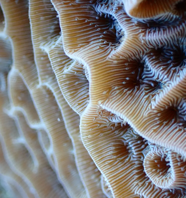
This is the intricate skeleton of a coral animal. Each polyp builds a tiny structure (corallite), forming a maze that helps me identify the species. Here, the polyps of Agaricia agaricites peek out from their skeletons, reaching into the water column in search of food.
The image shows the living coral skeleton and tissue of an Agaricia agaricites colony in St. John, US Virgin Islands. A. agaricites is one of the few species in this genus that are recognizable by their distinct skeleton. This photo shows the living coral tissue reaching out between to feed during the day. The color seen is from the symbiotic algae which live in the tissues of the coral to help supplement their food supply. This is a very important relationship, and one of the most famous examples of symbiosis in the animal kingdom. I took this image with an Olympus TG-6 camera, in about 15 feet of water, on SCUBA while conducting research surveys identifying species of coral in St. John, and how they might be responding to changes to habitat structure brought on by climate change.