Cardiac Rainbow
Description
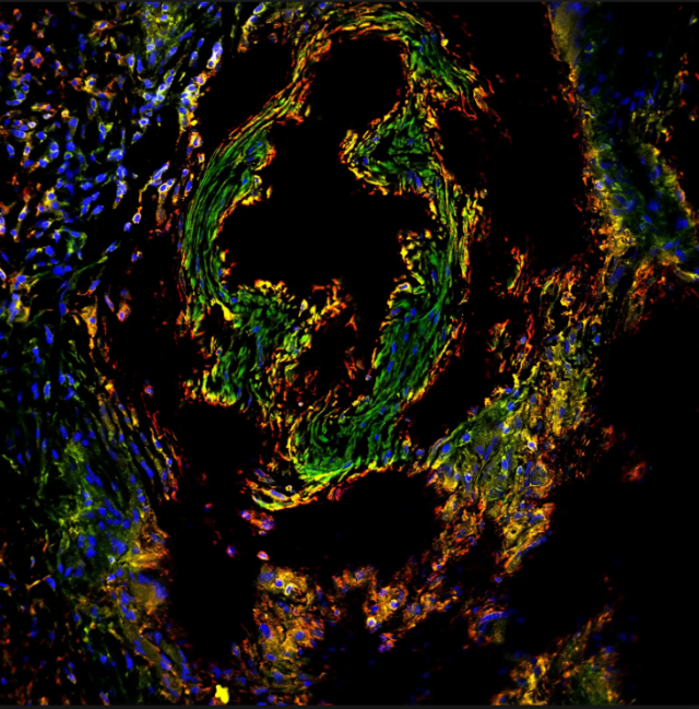
For this experiment, we wanted to understand how heart disease changes heart cell populations. To do this, we cut diseased human heart tissue into thin slices and stained cells with different fluorescent colors. Green cells are heart cells, and red cells are fibroblasts, or cells that are known to contribute to the disease. Yellow stains the outside of cells, and blue stains the nuclei of cells. Understanding how the types of cells, and what they look like, change during heart disease will allow us to better understand how heart disease works and come up with new and effective ways to treat it.
Image is a confocal image of an immunohistochemistry stain on human cardiac tissue. Tissue was stained with DAPI (blue), Phalloidin (yellow), alpha-actinin (green heart cells), and alpha- smooth muscle actin (red fibroblasts).