The World of a Cell
Description
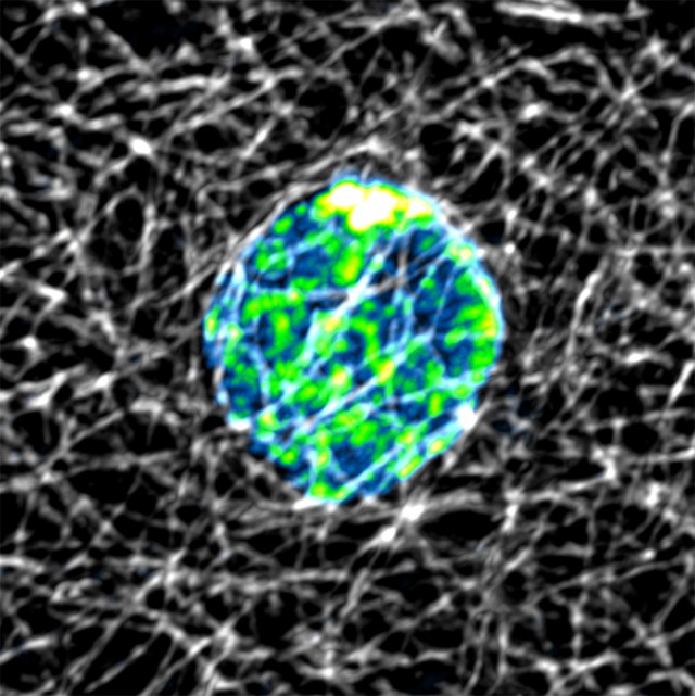
The World of a Cell, like the cosmos, are fascinatingly vast. Pictured is a cell from a fruit fly ovary, showing a central nucleus, the home of the genome. Surrounding it is a beautifully complicated meshwork of microtubule scaffolds that provide structure, rigidity and highways to traffic proteins around the cell. The nucleus of this cell looks like our own planet Earth, a metaphor to our larger, and smaller, place in the universe.
ZOOMING IN This projected confocal image is of a stretch cell in the ovary of the fruit fly, Drosophila melanogaster. Stretch cells surround the developing egg and exhibit a flat, or squamous, morphology. The earthly looking nucleus is highlighted by the small fluorescent DNA stain, Hoescht33342. The DNA in the nucleus is pseudocolored for green to blue based on variation in intensity, which shows that DNA is packed in patches within the nucleus. Gray microtubules that resemble cosmic rays are labeled using a technique call immunohistochemistry using fluorescent antibodies. This wonderfully complicated meshwork of tubules serves as a structural component, is a highway to traffic proteins around the cell, and is required to distribute DNA equally between two cells during cell division.


