Tensellation
Description
Cells make up every living thing found in nature, but no one really knows why they do the things they do. By looking at the forces that cells apply on each other, we can then make connections between applied forces and cellular movements.
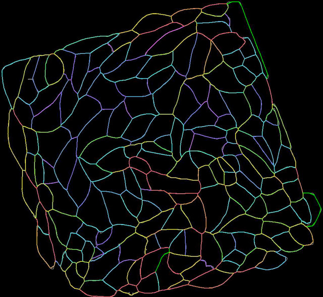
The above image is a “heat map” showing relative tensions felt by individual cells in a kidney tissue; red correlates with high tension and purple correlates with low. The key principle here is that tension states can be predicted from cellular geometry. Images of the original tissue were taken using fluorescence microscopy, and the cells were then individually outlined using a machine-learning based segmentation software. With a little bit more image analysis, the tension states were then extracted and displayed in the form of a color gradient. This is a highly efficient method of collecting and showcasing data, as it is a non-invasive procedure and immediately shows trends present in a tissue.


