The Microfluidic Blood-Brain Barrier
Year:
2017Ranking:
EntrantArtist:
Max Nowak (Graduate Student), Tyler Brown Department:
Chemical EngineeringLab:
Mitragotri/Helgeson LabsDescription
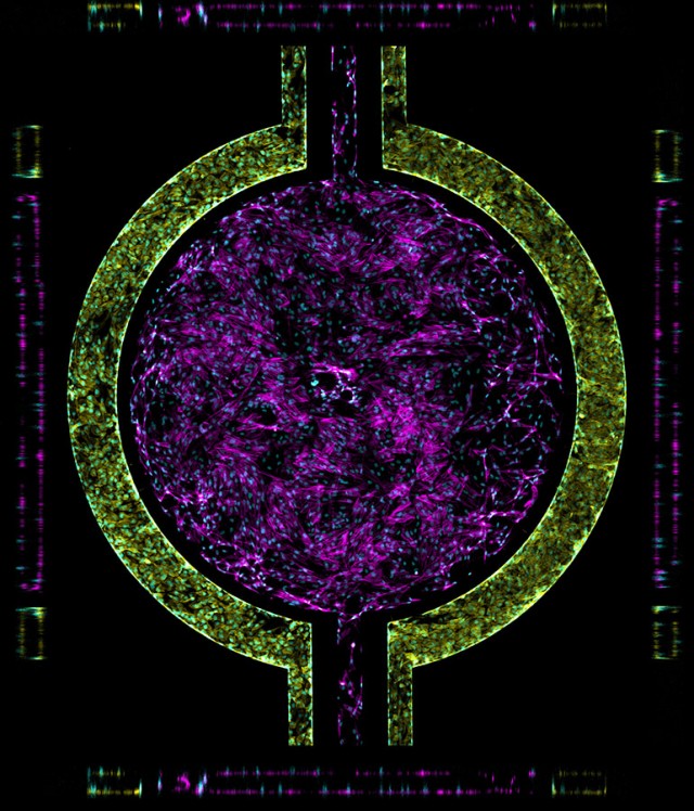
Top and side views of a microfluidic blood-brain barrier (BBB). Endothelial cells (yellow) line the side channel, forming the "barrier" part of the BBB while astrocytes (magenta) represent the "brain". Cyan spots are the nuclei of each individual cell.
Cells were grown in the microfluidic, then fixed and stained for actin. Image was collected via fluorescent confocal microscopy; Z-stacks with 30 slices each were taken for 25 different sections of the device and then stitched together. Image was enhanced slightly to aid visualization.


