A Fluorescent Retina
Year:
2016Ranking:
EntrantArtist:
Amanda Kautzman (Graduate Student)Lab:
Reese LabDescription
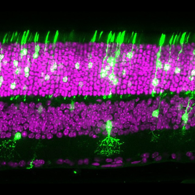
This is a cross section of an adult mouse retina. Retinal layers can be identified by nuclei stained with the dye DAPI (magenta). A subset of neurons were genetically manipulated to express a green fluorescent protein (green) in order to examine mechanisms of central nervous system development.
Cells were made to fluoresce green through a molecular biology technique called in vivo electroporation. In this technique, DNA is surgically injected into the eyes of newborn mice and following a delivery of a short electrical pulses, leads to the expression of foreign genes and proteins. The image was acquired on a confocal microscope and is taken at a 40X magnification.