Blood Cell Flow in Embryonic Zebrafish
Description
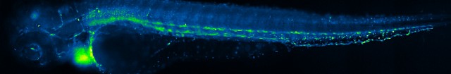
A lateral view of a 5 day old zebrafish under widefield fluorescence microscopy. In this particular zebrafish, Tg(gata1:dsRed) x Gt(desma:mCherry), red blood cells (the bright dots in the image) are fluorescent. During imaging, the zebrafish is kept alive (but immobilized by anesthesia) so we can observe the dynamics of blood flow and heart pumping. This image is a single frame from a movie created by stitching together ten 512x512 movies acquired at 30 fps and 20x magnification. Each individual movie was taken of a small region of the fish, one at a time, and they were all combined with 3D stitching in ImageJ to create a full lateral view (similar to stitching together a panorama). The image was also resized and colored with a green-blue lookup table in ImageJ.